Article Text

Statistics from Altmetric.com
Pineoblastomas occur in RB-1 mutation carriers,1 but are rarely seen in association with other hereditary syndromes.2 Germline DICER1 mutations are associated with embryonal tumours such as pleuropulmonary blastoma (PPB),3 ,4 so we hypothesised that such mutations might also occur in children with pineoblastoma. Here we describe a novel deleterious germline DICER1 mutation in a young child with a highly aggressive pineoblastoma. In addition, we observed loss of heterozygosity (LOH) of the wild-type allele, an event not previously reported in DICER1 associated tumours.5 ,6 Our observations suggest: (1) pineoblastomas are associated with germline DICER1 mutations; and (2) unexpectedly, tumour cells can thrive in the absence of functional DICER1.
The pineal gland, so named by Galen because of its resemblance to a pine nut, is a ∼175 mg midline structure situated on the posterior wall of the third ventricle, deep between the cerebral hemispheres. Pineal gland tumours are often derived from ectopic germ cell tissue, but pineocytomas and pineoblastomas also occur. A pineoblastoma is a supratentorial primitive neuroectodermal tumour arising in the pineal gland. Germline mutations in tumour suppressor genes have rarely been associated with pineoblastomas,1 ,2 but mutations in RB-1 can result in so-called ‘trilateral retinoblastoma’.1
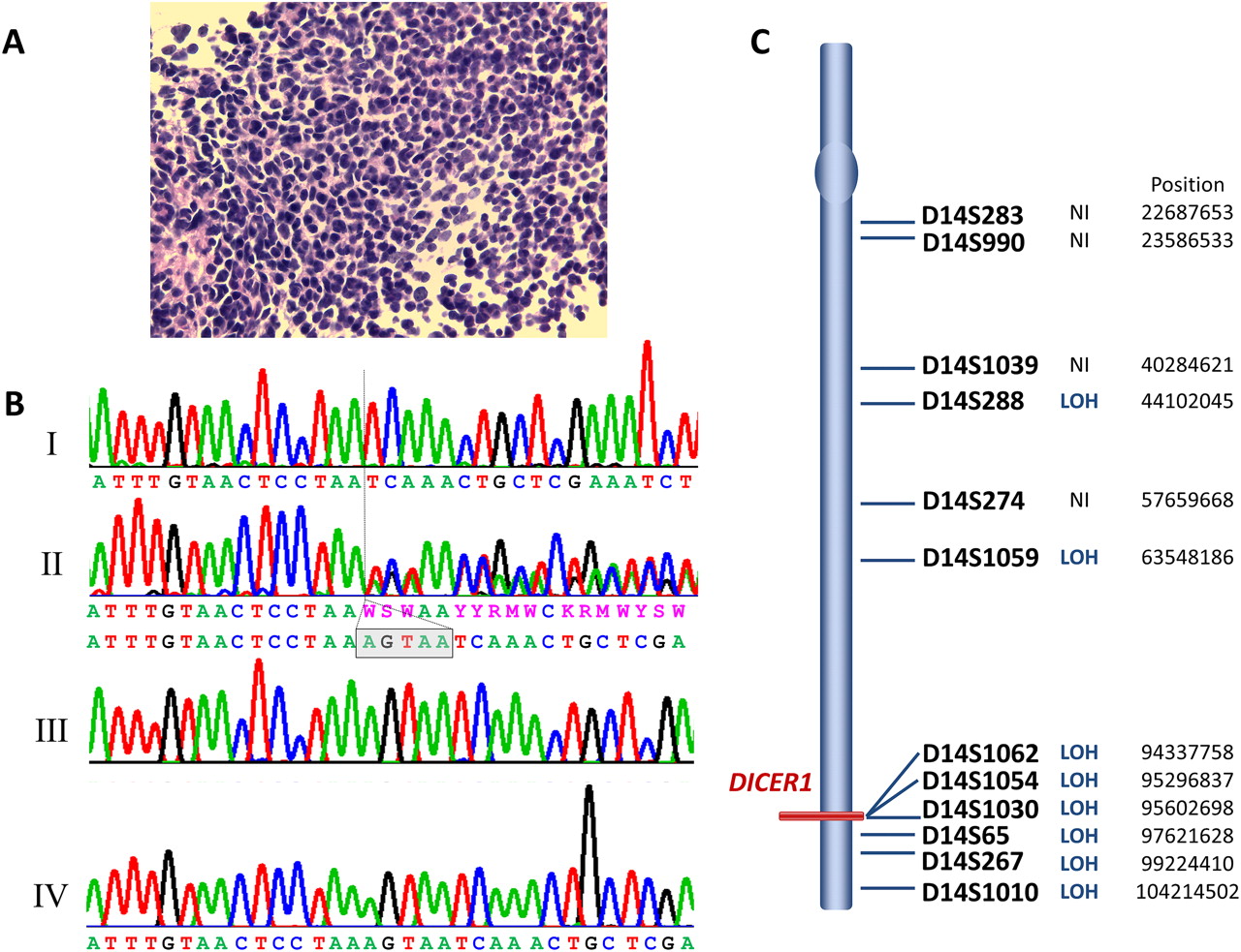
We obtained fresh-frozen tissue from a highly malignant, undifferentiated tumour occurring in a young child. The tumour consisted of sheets of ‘small blue’ cells with hyperchromatic nuclei and scant cytoplasm. Focally, they were surrounded by finely eosinophilic neuropil-like matrix. Definite Homer Wright rosettes, however, were not seen (figure 1A). Immunohistochemical staining was diffusely positive with antibodies directed against chromogranin, neuron-specific enolase and synaptophysin. The tumour was sparsely and focally positive for antibodies to glial fibrillar acid protein and neurofilaments (not shown). Electron microscopy revealed undifferentiated cells with scattered small intercellular junctions. The cells also formed small rosettes around minute lumens. The tumour cell cytoplasm was found to contain variable amounts of microtubules (not shown). In combination, these features confirm the diagnosis of a pineoblastoma. This sample was obtained from the Brain Tumour Tissue Bank (http://www.braintumour.ca/184/brain-tumour-tissue-bank). Under the terms of the establishment of the Bank, all samples are provided anonymously and therefore it is not possible to re-contact the family of the patient to attempt to gather further clinical information on the child or relatives.

(A) Haematoxylin and eosin stain of the pineoblastoma. The tumour features small blue cells with hyperchromatic nuclei and scant cytoplasm. Note the numerous mitotic figures. Original magnification ×400. (B) Chromatograms showing loss of heterozygosity of the wild-type allele in the pineoblastoma (panel I) and the heterozygous c.1128_1132delAGTAA mutation in the patient's blood (panel II), compared to the wild-type sequence (panel III). Sequencing of cDNA from the patient's blood showed wild-type sequence only (panel IV). The tumour is clearly hemizygous for the c.1128_1132delAGTAA allele, with no apparent contribution from the wild-type allele. The lack of mutant sequence in lymphocyte derived cDNA is presumably due to the presence of nonsense mediated decay. (C) Graphic representation of chromosome 14 showing the 12 microsatellite (STR) markers genotyped along with their loss of heterozygosity status. LOH, loss of heterozygosity in tumour DNA compared to blood DNA; NI, marker not informative (homozygous in the blood DNA); Position, approximate position of the marker on the chromosome.
We sequenced the coding exons of the DICER1 gene using genomic DNA isolated from this tumour and we identified a deleterious mutation in exon 8, c.1128_1132delAGTAA, which is predicted to result in p.Lys376Asnfs*11 at the protein level. Sequencing of whole blood-derived DNA from the same patient revealed that this mutation is present in the heterozygous state in the germline, but is hemizygous in the tumour (figure 1B, chromatograms II and I, respectively). Notably, sequencing of cDNA synthesised from RNA extracted from the blood sample showed no evidence of the five base pair exon 8 deletion (figure 1B, chromatogram IV), presumably because of the occurrence of nonsense mediated decay in the presence of a premature stop codon, resulting from the frameshift.
We genotyped 12 microsatellite markers to estimate the extent of the LOH. Eight markers were heterozygous in blood and thus informative (figure 1C). These markers span a region of 60 megabases on 14q, and all showed loss of one allele in the tumour. Based on these results, loss of the entire 14q arm bearing the wild-type DICER1 allele appears likely.
LOH at the DICER1 locus in the tumour, together with the frameshift mutation in the germline, indicates that, contrary to expectations from both animal models7 ,8 and data in humans,5 ,6 complete loss of functional Dicer1 protein can occur in human tumours. In animal models, in contrast to the effect of loss of one functioning allele, total loss of Dicer1 protein retards or prevents tumour development, suggesting that there is active selection against complete Dicer1 loss in tumours.7 ,8 In addition, LOH of the presumed wild-type allele was not observed in 28 human tumours bearing germline or somatic DICER1 mutations.3–6 ,9
One reason for the phenomenon of LOH might be that the very aggressive nature of this pineoblastoma (in contrast to many of the tumours arising in DICER1 mutation carriers) resulted in secondary events, including loss of the DICER1 locus, and by this time, the tumour had acquired sufficient genomic rearrangements to forego the apparent need for Dicer1 function. On the other hand, PPB can also be a very aggressive tumour. Until LOH studies are related to outcome (eg, one could study brain metastases from children with PPB), it will be impossible to know if the presence of LOH is in some way related to tumour aggressiveness.
Whether or not there is a link between LOH and tumour aggressiveness, this solitary finding refutes the notion that LOH of the wild-type allele, in the setting of a deleterious germline (or somatic) mutation on the other allele, is incompatible with tumour development. Unlike the recently reported existence of recurrent somatic DICER1 hotspot mutations in non-epithelial ovarian cancers (particularly Sertoli-Leydig cell tumours),6 the findings reported here are more compatible with a traditional ‘two-hit’ model of tumourigenesis, wherein complete loss of Dicer1 protein provides a selective advantage to tumour cells that have undergone this change.
Perhaps the spatio-temporal timing of DICER1 loss is also relevant; in some nascent tumour cells, early complete loss of Dicer1 is fatal to their development, whereas in others, later loss of Dicer1 function can be an advantage. These speculations can be addressed by further studies using other tumours associated with both germline and somatic DICER1 mutations.
Several ‘blastoma’-type tumours, most notably PPB, arrhenoblastoma (now known as Sertoli-Leydig cell tumour), embryonal rhabdomyosarcoma, and Wilms tumour (nephroblastoma), are associated with germline mutations in DICER1.3–5 ,9 A pineoblastoma has co-occurred in a child with an intraocular medulloepithelioma,10 and a mutation in DICER1 has been found in another child with this latter tumour.4 Thus, it appears that pineoblastomas fall within the expanding spectrum9 of tumours attributable to germline DICER1 mutations.
The relatively low penetrance of many DICER1 mutations suggests that mutation analysis of DICER1 in children with rare tumours such as pineoblastomas may be justified even in the absence of a family history of other cancers and related disorders. This could lead to the identification of unaffected DICER1 mutation carriers, which may permit early diagnosis of potentially fatal associated conditions, especially PPB.
Acknowledgments
We thank the Brain Tumour Tissue Bank, funded by Brain Tumour Foundation of Canada, for supplying the samples.
Footnotes
Funding The work was funded by the Mendon F Schutt Foundation.
Competing interests None.
Ethics approval Ethics approval was approved by McGill University.
Provenance and peer review Not commissioned; externally peer reviewed.