Article Text

Abstract
Background MicroRNAs (miRNAs) are non-coding gene transcripts involved in post-transcriptional regulation of genes. Recent studies identified miRNAs as important regulators of learning and memory in model organisms. So far, no mutations in specific miRNA genes have been associated with impaired cognitive functions.
Methods and results In three sibs and two unrelated patients with intellectual disability (ID), overlapping 1p21.3 deletions were detected by genome-wide array analysis. The shortest region of overlap included dihydropyrimidine dehydrogenase (DPYD) and microRNA 137 (MIR137). DPYD is involved in autosomal recessive dihydropyrimidine dehydrogenase deficiency. Hemizygous DPYD deletions were previously suggested to contribute to a phenotype with autism spectrum disorder and speech delay. Interestingly, the mature microRNA transcript microRNA-137 (miR-137) was recently shown to be involved in modulating neurogenesis in adult murine neuronal stem cells. Therefore, this study investigated the possible involvement of MIR137 in the 1p21.3-deletion phenotype. The patients displayed a significantly decreased expression of both precursor and mature miR-137 levels, as well as significantly increased expression of the validated downstream targets microphthalmia-associated transcription factor (MITF) and Enhancer of Zeste, Drosophila, Homologue 2 (EZH2), and the newly identified target Kruppel-like factor 4 (KLF4). The study also demonstrated significant enrichment of miR-137 at the synapses of cortical and hippocampal neurons, suggesting a role of miR-137 in regulating local synaptic protein synthesis machinery.
Conclusions This study showed that dosage effects of MIR137 are associated with 1p21.3 microdeletions and may therefore contribute to the ID phenotype in patients with deletions harbouring this miRNA. A local effect at the synapse might be responsible.
- MIR137
- microRNA-137
- 1p21.3 microdeletion
- intellectual disability
- synapse
- genetics
- microrna
- neurosciences
- chromosomal
- copy-number
- genetic screening/counselling
- diagnostics
- cytogenetics
- clinical genetics
- academic medicine
- neuromuscular disease
- memory disorders
- molecular genetics
- hydrocephalus
Statistics from Altmetric.com
- MIR137
- microRNA-137
- 1p21.3 microdeletion
- intellectual disability
- synapse
- genetics
- microrna
- neurosciences
- chromosomal
- copy-number
- genetic screening/counselling
- diagnostics
- cytogenetics
- clinical genetics
- academic medicine
- neuromuscular disease
- memory disorders
- molecular genetics
- hydrocephalus
Introduction
microRNAs (miRNAs) are small RNAs that are highly conserved and involved in several biological processes such as developmental timing, cell proliferation, apoptosis, metabolism, cell differentiation, and morphogenesis.1 They regulate gene expression by binding to the 3′-untranslated region (UTR) of target mRNAs and inhibiting protein synthesis or causing mRNA degradation.2 3 Each miRNA can act on many mRNAs and, conversely, individual mRNAs can be targeted by a number of miRNAs. Recent estimates puts the number of human miRNAs at 1100 or more, influencing the expression of as many as two-thirds of all genes.4 5 A few miRNAs—for example, miR-124, miR-134, and miR-338—are specifically expressed in brain, suggesting unique regulatory roles in neuronal development and function.5 6 The large number of miRNAs imposes a formidable challenge for studying miRNAs, their target mRNAs, and their functions in learning and memory, brain development, and brain plasticity.7 Multiple lines of evidence suggest that altered neuronal plasticity and morphology as seen in neurodevelopmental disorders may result from disruption of a common post-translational process that is under tight regulation by miRNAs.5 7 Several intellectual disability (ID) syndromes, such as fragile X syndrome, Rett syndrome, and Down syndrome, have been associated with miRNA pathways.8–12 Little is known, however, about the pattern of expression of these small non-coding RNA molecules in the brains of normally developing individuals or patients with disorders of the nervous system. Moreover, no previous reports have linked the disruption of specific microRNAs to ID.
While interstitial deletions of the short arm of chromosome 1 are rarely reported, a recently published study identified hemizygous microdeletions restricted to the chromosomal region 1p21.3.13 The individuals described had autism spectrum disorder and severe speech delay. The authors of this study suggested that hemizygous deletions of the dihydropyrimidine dehydrogenase (DPYD) locus are contributing to these phenotypes. Though absence of reports of abnormal cognitive functioning in the many heterozygous carriers of DPYD loss-of-function mutations goes against an exclusive role for DPYD in the observed cognitive impairment,14 DPYD is involved in autosomal recessive dihydropyrimidine dehydrogenase (DPD) deficiency. The phenotype of DPD deficiency is variable, but frequently comprises developmental delay and convulsions.15
The present study involves five individuals with ID who have overlapping 1p21.3 microdeletions that included DPYD but also the miRNA gene microRNA 137 (MIR137). Expression studies revealed reduced levels of precursor and mature microRNA-137 (miR-137) and increased levels of downstream targets of miR-137 in the patients compared to controls. Our studies indicate that alteration in miR-137 expression levels is a likely explanation for their ID phenotype. To our knowledge, this is the first study to demonstrate that a reduced copy number of a specific miRNA might be associated with ID.
Patients and methods
Patients
Patients 1, 2 and 4 were ascertained by genome-wide array analysis during a pending cohort study in patients with unexplained ID at the department of human genetics in Nijmegen, The Netherlands. Patient 5 was ascertained by genome-wide array analysis during routine diagnostic evaluation of her ID at the departments of clinical genetics and neuropediatrics in Lille, France. Patients 1–3 were siblings. From patient 3 we obtained a blood sample for genetic testing and concise information on her developmental level, but it was not possible to perform a clinical examination. Chromosomal analysis in the parents of the three affected sibs was not possible because they were deceased. They were known to have low cognitive abilities and had not been able to take care of their children, who were placed in foster homes. Patients 4 and 5 were single unrelated patients. Fragile X syndrome and Prader–Willi syndrome were excluded in patients 1 and 2 from family 1 and in patients 4 and 5.
Single nucleotide polymorphism array analysis
DNA was obtained from peripheral blood leucocytes and isolated according to standard procedures. In patients 1–4, as well as in the parents of patient 4, genome-wide single nucleotide polymorphism (SNP) array analysis was performed by the Affymetrix 250k SNP array platform according to the standard Affymetrix GeneChip protocol (Affymetrix, Inc, Santa Clara, California, USA). In patient 5 and her parents genome-wide array analysis was performed by the Agilent 44 k array platform. Copy number estimates were determined using the updated version 2.0 of the CNAG (Copy Number Analyzer for Affymetrix GeneChip mapping) software package.16 The normalised rations were subsequently analysed for genomic imbalances by a standard Hidden Marker Model (HMM). Copy number variations (CNVs) were mapped according to the USCS genome browser build March 2006. To confirm the deletions detected by 250 k SNP array analysis, quantitative PCR (qPCR) analysis on DNA extracted from blood lymphocytes was performed.
Metabolic tests
In patients 1 and 2 (siblings) and in patient 4, measurements of serum and urine concentrations of purines and pyrimidines were performed to rule out DPD deficiency.
RNA isolation
Total RNA (1–5 μg) was isolated from cell suspensions of approximately 1×10⁶ Epstein–Barr virus (EBV) transformed B cells from individual cell lines using the NucleoSpin RNA II RNA isolation kit form Machery–Nagel (Düren, Germany). The concentration and quantity of RNA were measured by ultraviolet (UV) absorbance at 260 and 280 nm (A260/280 ratio) and checked by gel electrophoresis.
Quantitative reverse transcription PCR of precursor and mature miRNA-137 and its downstream targets
Quantitative reverse transcription PCR (qRT-PCR) quantification of precursor and mature miR-137 levels and its validated and newly identified downstream targets was performed on RNA from lymphoblastoid cell lines (LCL) of patients and controls, as well as on RNA from several human brain regions and mouse cortical synaptosomal preparations, according to previously described protocols.17 18 Precursor miR-137 primers were designed to anneal within the hairpin sequence of the miRNA-137 precursors. Precursor miRNA-137 cDNA was synthesised from total RNA by using gene specific miR-137 reverse PCR primers. The forward primers for qPCR analyses for mature miR-137 were designed using the entire mature miRNA sequence (primer sequences are shown in table 1).
Primers used for quantitative reverse transcriptase PCR of precursor and mature miR-137
Mature miR-137 cDNA was synthesised from total RNA (1 μg) using the NCode miRNA First Strand cDNA synthesis kit (Invitrogen, Carlsbad, California, USA) according to the manufacturer's protocol. Using first-strand cDNA as a template, triplicate qPCR reactions were performed from samples derived from three different experiments for mature miRNA using miR-137 specific forward primers and the NCode Universal qPCR reverse primer. Minus template and minus forward primer controls were included to ensure lack of signal in the assay background. U6 snRNA levels were used as a loading control. All CT (threshold cycle) values used for analyses were averaged from three to six replicates and those with high SD (>1) were not included in the analyses. Melting curve analysis was performed to test for the specificity and quality of the qPCR amplifications. Relative expression was calculated using the comparative CT method18 normalised to the expression of U6 snRNA. To assess the levels of miR-137 downstream target mRNAs, 2 μg of DNAse-treated, total RNA from each sample was used for cDNA synthesis, using the RevertAid H Minus First Strand cDNA Synthesis kit (Fermentas Inc, St. Leon-Rot, Germany). Before qPCR analysis, each cDNA sample was diluted 1:10 with MilliQ water. qPCR was performed according to previously described protocols,17 18 using standard cycling conditions and performing a melting protocol to control for product specificity. The Maxima SYBR Green qPCR Master Mix Kit (Fermentas Inc, St. Leon-Rot, Germany) was used in the qPCR reactions. Primers were designed using NCBI Primer-Blast (http://www.ncbi.nlm.nih.gov/tools/primer-blast/) and synthesised at Biolegio BV (The Netherlands). Primer sequences for MITF, EZH2, and KLF4 are available upon request. The relative levels of the transcripts of the downstream targets were normalised to β-glucuronidase (GUSB) and β-actin (ACTB) mRNAs to provide an internal control for reverse transcription. These messages were used as reference genes because of their stable expression in lymphocytes.19 All other human RNAs investigated were purchased from Stratagene (La Jolla, California, USA).
Synaptosomal preparation
Synaptosomes were isolated based on the method of Gray and Whittaker.20 All the procedures were performed at 4°C, and under RNase-free conditions. Briefly, mouse cortical tissue was homogenated using a Dounce Homogenizer (B Braun Biotech International, Melsungen, Germany) in 5 mM HEPES buffer containing 0.32 M sucrose, protease inhibitor cocktail (Roche, Basel, Switzerland) and 1 U/ml RNase inhibitor (Fermentas, St. Leon-Rot, Germany). The homogenate was centrifuged at 1000 g for 10 min, obtaining supernatant S1 and pellet P1. Pellet P1 was resuspended with homogenisation buffer and centrifuged at 1000 g for 10 min, obtaining supernatant S2 and pellet P2 (containing the nuclear fraction). Supernatants S1 and S2 were pooled together and centrifuged at 20 000 g for 10 min, obtaining supernatant S3 (containing the cytoplasmic fraction) and pellet P3. Pellet P3 was resuspended in homogenisation buffer, loaded on a discontinuous sucrose density gradient (0.85 M and 1.2 M sucrose layers), and centrifuged at 30 000 g for 2 h. The interface between the two sucrose layers was collected and centrifuged at 25 000 g for 30 min, obtaining a pellet containing the synaptosomal fraction. The purity of the synaptosomal fraction was determined by western blot assay using the synaptic marker (PSD95, 1:5000; NeuroMab, Davis, CA, USA) and markers known not to be enriched at the synapse (tubulin, 1:500; in-house antibody). GADPH (1:1000; Cell Signaling, Boston, MA, USA) was used as a housekeeping marker. The nuclear, cytoplasmic and synaptosomal fractions were kept at −80°C until further analysis. RNA from nuclear, cytoplasmic and synaptosomal fractions was isolated with TRIzol Reagent (Invitrogen), according to the manufacturer's protocol. The procedure was modified for small amounts of tissue by using 800 μl of TRIzol Reagent and adding 1 μl of glycogen (Fermentas, St. Leon-Rot, Germany). RNA concentration and quality was determined with a NanodropTM ND-1000 spectrophotometer (Thermo Fisher Scientific, Lafayette, CO, USA) and 1% agarose gel electrophoresis, respectively. The samples were kept at −80°C until further analysis.
miRNA computational predictions
To assess the potential regulatory impact of miR-137 at the synapse, we searched the TargetScan miRNA target database for conserved, putative rodent targets of miR-137.21 22
Neuronal cell cultures and transfections
Primary cultures of cortical neurons were prepared from embryonic day 18 rats as described23 and maintained in a neurobasal medium supplemented with B27 (Invitrogen) and 2 mmol/l glutamine. For miR-137 expression experiments, the miRNASelect pEGP-mmu-mir-137 Expression Vector (Cell Biolabs, Inc, San Diego, California, USA) constructs expressing mouse miR-137 precursor and GFP, or the negative control pEGP-null vector (expressing GFP), were purchased. Locked nucleic acid miR inhibitor, anti-miR-137, as well as non-targeting control anti-miR-NT were obtained from Exiqon (Denmark). Small RNAs and Constructs were introduced into primary neurons using Lipofectamine 2000 (Invitrogen).
Statistical analysis
Student t test was used to determine significant differences in transcript levels between the patients and the controls.
One-way analysis of variance (ANOVA) was used to analyse significant differences among multiple groups; p<0.05 was considered significant.
Results
Clinical results
A summary of the clinical features is shown in table 2.
Clinical and molecular features of overlapping 1p21.3 microdeletions in patients 1–5
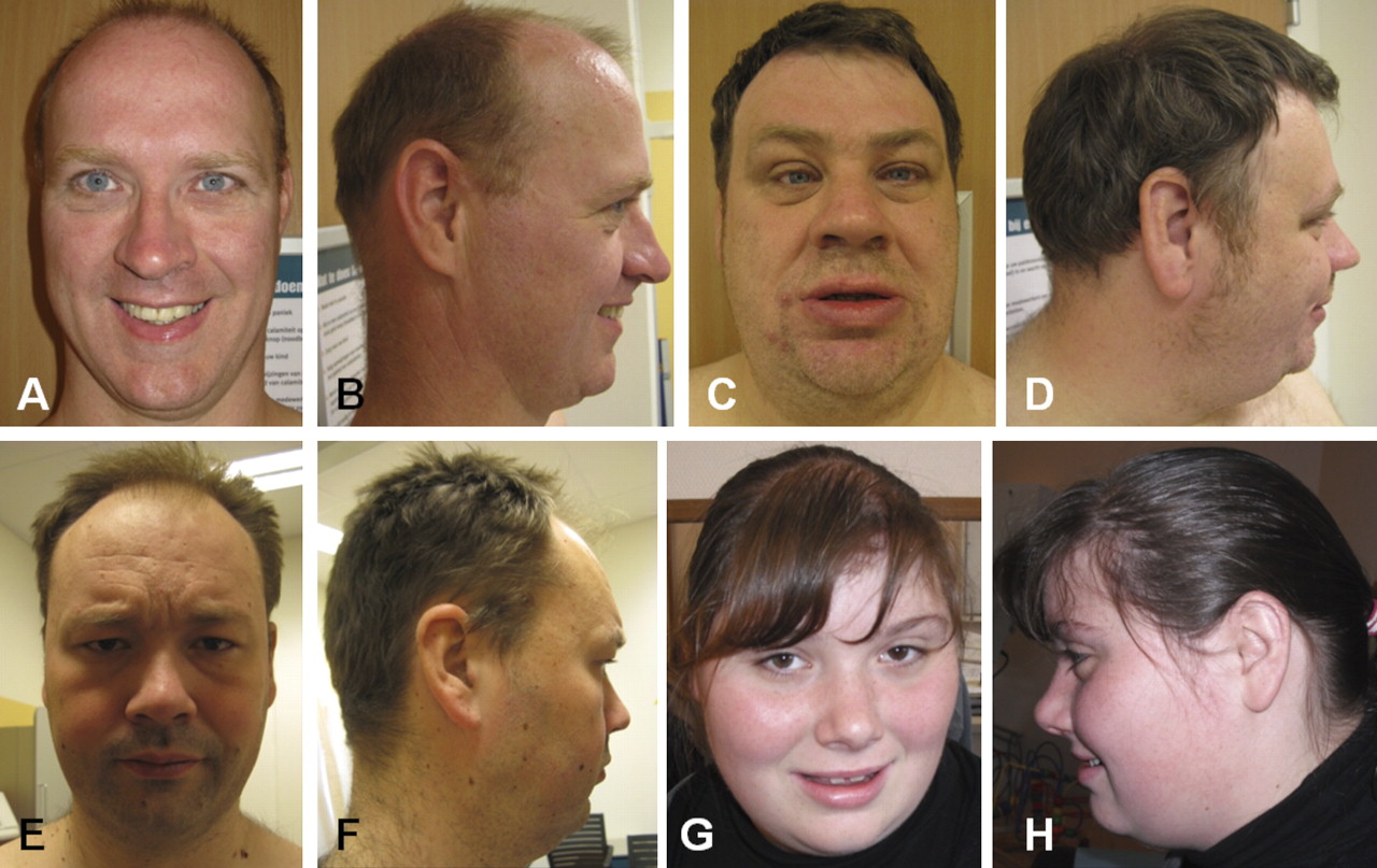
Photographs of patients 1, 2, 4, and 5 are shown in figure 1. Though the patients have no distinct dysmorphic facial features, they showed similarities in facial appearance, including long ears, deeply set eyes, broad nasal tip, and thick lower lip. The level of ID ranged from borderline-mild (patient 1) to mild-moderate (patients 2 and 3). Remarkably, patients 1, 2, and 4 showed a similar discrepancy between verbal and performal IQ with a relatively low score on verbal capacities. Such a discrepancy was not reported in patient 5. Patient 2 of family 1 and patient 4 also showed similar speech deficits including poor intelligibility and specific pronunciation deficits (in patients 1, 3, and 5 speech tests were not performed). Similar behaviour characteristics included a remarkable shy and friendly behaviour, (tendency) to overeating, and features of autism spectrum disorder. Patients 1, 2, 4, and 5 were obese or had a tendency to obesity. Patients 1, 2, and 4 had non-specific ocular problems.

Facial appearance of patients 1, 2, 4, and 5. Panels A and B show patient 1 at age 42 years, panels C and D patient 2 at age 38 years, panels E and F patient 4 at 34 years, and panels G and H patient 5 at age 18 years. Note the minor facial dysmorphic features, including long ears, deeply set eyes, full tip of the nose, and thick lower lip.
Serum and urine measurements of purine and pyrimidine levels in patients 1, 2, and 4 revealed no abnormalities indicative for DPD deficiency.
Genome-wide array analysis
Figure 2 provides a schematic representation of the overlapping deletions in 1p21.3 in our patients and in the patients reported by Carter and others.13 In patients 1–3 an identical interstitial 1.75 Mb loss was identified (97, 50–99, 25 Mb, according to Hg 18 UCSC genome browser): 46, XY/XX.arr snp 1p21.3 (SNP_A-2211294→SNP_A-2131140)×1. In patient 4 an interstitial loss of 1.41 Mb was identified (97, 32–98, 73 Mb, according to Hg 18 UCSC genome browser): 46,XY, 1p21.3 (SNP_A-1881473→SNP_A-1789641)×1. In patient 5 an interstitial loss of 2.45 Mb was identified (96, 27–98, 72 Mb, according to Hg 18 UCSC genome browser): 46, XX, 1p21.3 (probe A_14_P100140→probe A_14_P113179). The deletion of patients 1–3 comprised three annotated Refseq genes, including DPYD, SNX7, and LPPR5, and the microRNA gene MIR137. The deletion of patient 4 comprised the annotated gene DPYD and MIR137 and the deletion of patient 5 comprised the annotated genes PTBP2 and DPYD and MIR137. The shortest region of overlap is 1.22 Mb in size and included DPYD and MIR137.

Schematic representation of the overlapping deleted regions in chromosome 1p21.3 identified by genome-wide array analysis. The small box indicates the chromosomal location that is schematically depicted below. The relative positions of the deleted regions in patients 1–3, 4 and 5 are indicated by black bars. The deletions in the patients reported by Carter and others12 are indicated in grey. The relative positions of the genes PTBP2, DPYD, SNX7, LPPR5, and MIR137 are indicated. The deletion of patient 5 extends distally beyond the figure. SRO, shortest region of overlap (in the present patients).
Genome-wide array analysis in the parents of patient 4 and 5 showed that the deletions in these patients had occurred de novo. DNA samples from the parents of patients 1–3 were not available for segregation analysis because they were deceased.
Differential miR-137 expression in LCL from the patients
To study the effect of loss of one copy of MIR137, we performed qRT-PCR on precursor and mature miR-137 in patient 2 (one of the siblings) and patient 4. LCL from patient 5 were not available. As controls we used RNA extracted from the LCL of three healthy male controls. Decreased precursor and mature miR-137 levels were found in the LCL from patient 2 as well as the LCL from patient 4, as compared to healthy controls and normalised to snoRNA U6 (one way ANOVA, p<0.001) (figure 3). The results demonstrate a great degree of reduction of miR-137 levels in the patients.

Precursor and mature miR-137 levels are decreased in the patients. The quantitative reverse transcriptase PCR assay was used to assess levels of precursor and mature miR-137 isolated from either the LCL of patients 2 and 4 and three healthy controls (HC). Levels of precursor (A) and mature (B) miR-137 levels are expressed relative to U6 snRNA which was used as an internal control. Error bars represent the SEM for triplicate PCR reactions from n=3 independent experiments; one way ANOVA, ***p<0.0001.
Increased levels of MITF and EZH2 in patient cell lines
While the major effect of miRNAs has been proven to be on the translation of target mRNAs, recent studies indicate that miRNAs also have a specific impact on the mRNA levels.3 24 We therefore investigated whether reduced levels of miR-137 in our patients corresponded to an increased level of the previously validated miR-137 targets EZH225 and MITF.26 To assess miR-137-mediated modulation of EZH2 and MITF mRNA levels, we monitored transcript levels in LCL of our patients and healthy controls by qRT-PCR. This investigation revealed significantly increased MITF and EZH2 levels in the patients as compared to two healthy controls (figure 4).

Validated miR-137 targets MITF and EZH2 mRNA levels are increased in the patients. The quantitative reverse transcriptase PCR assay was used to assess mRNA levels of MITF (A) and EZH2 (B) isolated from either the lymphoblastoid cell lines of patients 2 and 4, and two healthy controls. mRNA levels are expressed relative to β-actin and GusB which were used as an internal controls. Error bars represent the SEM for triplicate PCR reactions from n=3 independent experiments; one way ANOVA, *p<0.05.
miR-137 is enriched in the hippocampus and cortical brain regions, and highly abundant at the synaptodendritic compartments of neurons
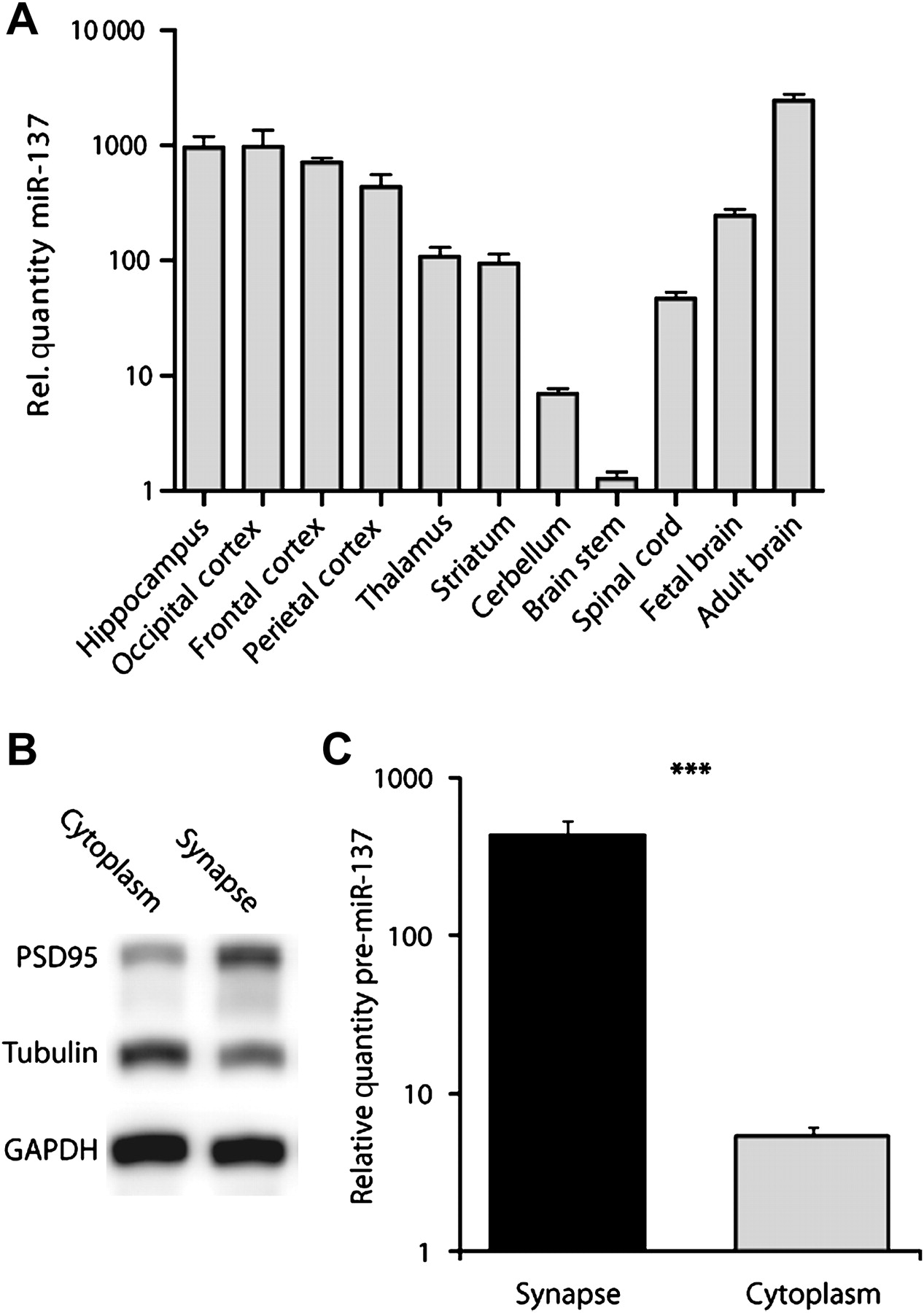
Many miRNAs have been isolated from mammalian embryonic neurons and mature brains and some of them are neuronal specific.27 Although miR-137 is specifically expressed in neuronal tissue,28 little is known about its abundance and function during neuronal maturation. To analyse the miR-137 level expression patterns in embryonic and mature brain, qRT-PCR was performed on total RNA isolated from fetal and adult (postmortem) human brain. As shown in figure 5A, miR-137 levels are approximately 100 times higher in the adult as compared to embryonic brain, suggesting that the expression of miR-137 is increasing during brain development and during neuronal differentiation, indicating that it might have an important role in human brain development and functioning.

miR-137 is overexpressed in the hippocampus and cortical brain regions, and enriched at synaptic compartments. (A) Differential brain regional distribution of miR-137. The quantitative reverse transcriptase PCR (qRT-PCR) miRNA assay was used to quantify precursor mir-137 across different human brain regions. (B) Differential fractionation was used to isolate mouse synaptosomal compartments, and the preparations were assessed by western blotting using PSD95, GAPDH, and tubulin antibodies. (C) qRT-PCR based quantification of precursor mir-137 isolated from the synaptosomal and the cytosolic fractions of mouse brain. Levels of precursor mir-137 are expressed relative to U6 which was used as an internal control. Error bars represent the SEM for triplicate PCR reactions from n=3 independent experiments; t test, ***p<0.0001.
Using commercially available RNA isolated from a subset of human brain compartments, we showed that miR-137 is most abundant in the hippocampus. Similarly, high miR-137 levels can be detected in the occipital, frontal, as well as the parietal cortical regions, while the lowest miR-137 levels were detected in the brain stem and cerebellum (figure 5A).
Recent findings indicate that miRNAs can be enriched in synaptic compartments of neurons.29 This notion suggests that miRNAs may be ideally positioned to quickly regulate local translation in response to synaptic activity.30 A recent study revealed that the neuron-enriched miR-137 has a prominent role in the synapse maturation and morphogenesis of young neurons.31 We therefore hypothesised that miRNA-137 might reside near the synapse, where it could act as local regulator of the translation of synaptically enriched target mRNAs. Recent studies employed biochemically isolated synaptosomes from young rat brains to demonstrate that these small vesicular structures preserve components of local protein synthesis, including polyribosomes, mRNAs and regulatory RNAs, including microRNAs.32 33 We used differential fractionation to isolate mouse synaptosomal compartments, and the preparations were assessed by western blotting using PSD95, GAPDH, and tubulin antibodies (figure 5B), as well as by real-time RT-PCR measurements of CAMK2a, to verify that these components were enriched relative to total forebrain homogenate. This experiment revealed significant enrichment of miR-137 in RNA preparations from mouse synaptosomes compared to the cytosolic fractions, as assessed by qRT-PCR (figure 5C). The outcome of this experiment suggests that miR-137 may have a local role in translation dependent synaptic morphology.
Regulation of neuronal levels of KLF4 mRNA by miR-137
To further assess the potential regulatory impact of miR-137, we searched the TargetScan database for additional mRNAs that contained a putative miR-137 binding site.34 This revealed a large number pf putative miR-137 target mRNAs (data not shown). Among these mRNAs are three members of the Kruppel-like factor genes—KLF4, KLF11, and KLF12—which contained highly conserved binding sites for miR-137 in their 3′UTRs. Kruppel-like factor proteins (KLFs) are a family of transcriptional repressors associated with axon growth in central nervous system (CNS) neurons,35 and previous studies identified coordinated activities of different KLFs to enhance the regenerative capacity of CNS neurons. The predicted miR-137 target site (MTS) in KLF4 3′UTR (figure 6A) had a high aggregate PCT score with the cognate miR (0.77), suggesting a conserved targeting of the MTS for miR-137.36

miR-137 target KLF4. (A) Putative miR-137 binding site in the rat Klf4 3′ UTR. The 6 nt seed sequence is marked by vertical lines between the nucleotides. (B) miR-137 over expression reduces Klf4 levels in rat primary cortical neurons. (C) Quantification of Klf4 mRNA levels in rat cortical neurons transfected with anti-miR-137, or non-targeting short oligonucleotides (anti-miR-NT), as determined by quantitative reverse transcriptase PCR 72 h after oligonucleotide transfection. (D) KLF4 mRNA levels are increased in patients 2 and 4. mRNA levels are expressed relative to β-actin which was used as an internal control. Error bars represent the SEM for triplicate PCR reactions from n=3 independent experiments; one way ANOVA, *p<0.05.
To explore whether miR-137 regulates Klf4 mRNA levels in neurons, we monitored Klf4 mRNA levels after transfecting rat cortical neurons (DIV 10) with a miR-137 precursor vector. miR-137 transfection with an expression vector resulted in a 1000-fold increase in mature miR-137 levels compared with the endogenous miR-137 levels in null vector transfected cortical neurons (not shown). Klf4 mRNA levels decreased 20% in miR-137 overexpressing cortical neurons when compared with the null-vector transfected neurons (figure 6B). Conversely, transfection of neurons with a locked nucleic acid inhibitor of miR-137 resulted in a threefold increase in Klf4 mRNA as early as 72 h after transfection (figure 6C), when compared with the non-targeting anti-miR control.
The results from the miR-137 overexpression or inhibition experiments in rat cortical neurons prompted us to investigate KLF4 levels in LCL from our patients. This revealed significantly increased KLF4 mRNA levels in the patients as compared to the healthy controls (figure 6D).
Discussion
We present evidence for likely involvement of MIR137 in the ID phenotype of patients with 1p21.3 microdeletions. The shortest region of overlap included DPYD and MIR137. DPYD is involved in autosomal recessive DPD deficiency, which is characterised by developmental delay and convulsions.15 Several arguments oppose an (exclusively) causative role of deletion of DPYD in the ID phenotype of the presented patients. First, none of the present patients in whom metabolic tests were performed (patients 1, 2, and 4) showed the typical thymine-uraciluria associated with DPD deficiency. Second, although heterozygous mutations in DPYD, including loss-of-function mutations, occur frequently and contribute to toxicity for the anti-cancer drug 5-fluoroacil (5-FU),14 ID has not been reported in heterozygous carriers. In a recent report, four patients from three families with overlapping microdeletions in the same region were described.13 It was suggested that the associated phenotype, consisting of speech delay and autism, was due to hemizygous deletion of DPYD. However, one of the deletions in this report also comprised MIR137, and the identical deletion reported in two siblings was located only 40 kb proximally from MIR137 and might affect expression of MIR137. Finally, the fourth patient had an intragenic DPYD deletion which was inherited from a healthy mother and thus likely represented an inherited variant similar to many heterozygous carriers that are known. It is likely that one of the parents of sibs 1–3 from the present report also had the deletion, but both parents had impaired cognitive abilities and were not able to raise their children who, consequently, had been placed in foster care. Therefore, we conclude that at least for the patients with a heterozygous deletion encompassing MIR137, the phenotype is more likely associated with haploinsufficiency of MIR137, although a role for DPYD is not fully excluded. Indeed, previous reports of miR-137 studies showed that its expression is epigenetically regulated by Methyl-CpG-binding protein 2 (MeCP2) during different aspects of neurogenesis in mice.25 31 Therefore, MIR137 represents a good candidate for the observed ID phenotype in our patients.
There is one other example of the involvement of microRNA mutations in a human genetic disorder: mutations in MIR96 cause autosomal dominant hearing impairment.37 In addition some reports have shown that disruption of the recognition sites in target genes for specific miRNAs might be associated with a genetic trait in humans. In this regard, some evidence was provided that a mutation in the binding domain for miR-189 in the SLITRK1 is associated with Tourette syndrome and other neuropsychiatric features.38 Other studies have highlighted the importance of miRNA pathways in neuronal processes. A recent study39 showed increased cognition of adult Dicer1 mutant mice, lacking miRNAs (such as miR-137) in mature neurons in adult brain compared to controls, thereby providing evidence for a role of miRNAs as key players in learning and memory processes of mammals. In addition, recent findings indicate that miRNAs may be involved in modulating short term synaptic plasticity.40 Furthermore, synaptic activity can influence miRNA expression: recent reports indicated that miRNA levels are altered in hippocampal neurons that are induced to display long term depression or long term potentiation.41 42 This finding suggests that different levels or variation in miRNA expression levels will affect translation of synaptic proteins and consequently synaptic plasticity. Expression studies of precursor and mature miR-137 in our patients and controls demonstrated significantly reduced expression levels in the patients. We performed these studies in LCL of the patients. A number of previous investigations assessed the relevance of using LCL as probes to study the genetics of neurodevelopmental disorders.43 44 Baron and others43 assessed the feasibility of using LCL for genome-wide expression profiling and showed the usefulness of this tool for identifying genes associated with neurodevelopmental disorders. As shown in figure 3, the expression of miR-137 is more than twofold reduced. This might be explained by the short half life of miR-137 in mammalian cells45 in combination with an elevated synthesis of proteins involved in RNA turnover processes which are targeted by miR-137 (caused by a reduced inhibition of these proteins due to a reduced level of miR-137). This could further accelerate RNA turnover in the patients, which would result in lower miR-137 levels.
Our studies in mice also suggested an enrichment of miR-137 in RNA preparations of mouse synaptosomes, corresponding with the results of a recent study that revealed a significant role of the neuron enriched miR-137 in the synapse maturation and morphogenesis of young neurons.31 This suggests that miR-137 may have a local role in translation dependent synaptic morphology and that defects in miR-137 could interfere with proper synaptic signal transduction, thereby affecting cognitive brain function. We also showed a significantly increased expression of the previously validated downstream targets MITF and EZH2 and the newly identified downstream target KLF4. MITF encodes a transcription factor that is involved in the regulation of differentiation and development of melanocytes in the retinal pigment epithelium. Heterozygous loss-of-function mutations in MITF are involved in Waardenburg syndrome type 2A (MIM 193510) and Tietz syndrome (MIM 193510). As expected, our patients, who have an opposite dosage effect of MITF, did not show the classic features of these syndromes (hypopigmentation and deafness).
The downstream target EZH2 is a histone H3 lysine 27 methyltransferase and a member of the Polycomb protein family. It functions as a transcriptional repressor. The protein regulates CpG methylation by direct interaction with DNA methyltransferases.46 Histone methylation plays an important role in modulating chromatin structure and function. It was previously shown that the EZH2 protein interacts with the ATR-X gene which is involved in α-thalassaemia/mental retardation syndrome (ATRX syndrome) (MIM 301040).47 Furthermore, several other genes that are associated with ID phenotypes, such as MECP248 and EHMT1,49 are involved in chromatin modulation. Therefore, an association between altered expression of the downstream target EZH2 with the ID phenotype in our patients might very well be possible. In addition, the newly identified downstream target KLF4 might contribute to the ID phenotype as well, since it was shown to act as a transcriptional repressor of axon growth in the CNS.35 The reduced expression of miR-137 likely results in significantly altered expression of other putative miR-137 targets as well. Moreover, the identification of additional aberrations in MIR137 in other patients with ID and normal genome-wide array results would provide further evidence for the role of MIR137. In our in-house database, comprising the results of genome-wide array analysis in more than 4000 ID patients and controls, no additional deletions restricted to 1p21.3 have been registered. In addition, we screened a large cohort of approximately 1000 ID patients, who had previously tested negative for fragile X and/or Prader–Willi syndrome and had normal karyotypes, for MIR137 aberrations. We selected this cohort of patients because no designated phenotypic features were present except ID and overweight. We have not found additional MIR137 aberrations (results not shown), suggesting that disruption of MIR137 would be a rare cause of ID.
To unravel the underlying mechanism further, additional expression studies of other known downstream targets that are expressed in the dendrites are warranted. Furthermore, future studies are needed to determine further the synaptic role of miR-137 as a regulator of local translation in dendrites which is known to be a pivotal mechanism of synaptic maturation and plasticity.
Acknowledgments
We thank the participating patients and parents.
References
Footnotes
AA and TK contributed equally to this work.
Funding This work was supported by grants from the Consortium ‘Stronger on your own feet’ (to TK, MCCH, MHW); the Donders Center for Neuroscience fellowship award of the Radboud University Nijmegen Medical Centre (to AA); the FP7-Marie Curie International Reintegartion Grant (to AA); GENCODYS, an EU FP7 large-scale integrating project grant (grant number 241995) and ZonMW (clinical fellowship, grant number 90700365 to TK).
Competing interests None.
Patient consent Obtained.
Ethics approval This study was approved by the local ethical committee of the Radboud University Nijmegen, The Netherlands.
Provenance and peer review Not commissioned; externally peer reviewed.