Article Text

Statistics from Altmetric.com
Spinal muscular atrophy (SMA) is the second most frequent autosomal recessive disease, with a prevalence of 1 in 6000 live born infants.1 It is characterised by degeneration of motor neurones of the anterior horn of the spinal cord, leading to symmetrical muscular weakness and atrophy. The International SMA Consortium classification2 defines several degrees of severity in the SMA phenotype, depending on the age of onset and motor development milestones. Type I SMA, Werdnig-Hoffmann I disease, is the most severe form with onset within 6 months of birth. Patients are unable to sit up and have serious respiratory dysfunction. Type II SMA is the intermediate form with onset within the first 2 years; children can sit up but are unable to walk. The clinical course is variable. Type III (also called Kugelberg-Welander disease) begins after 2 years of age and usually has a chronic evolution. Children can stand and walk unaided at least in infancy. Adult form (type IV) is the mildest, with onset after 30 years of age; few cases have been reported and its prevalence is not accurately known.
Spinal muscular atrophy is linked to locus 5q13 in more than 95% of patients.3–6 The critical region, containing several genes including the survival motor neurone (SMN) gene, is inverted and duplicated. Homozygous deletion of SMN1, located in the telomeric position, accounts for the disease in 98% of these cases and has been reported in infantile, intermediate, and adult onset disease.7–10 Linkage analysis in families with SMA shows large de novo deletions in 2% of patients.11–13SMN2 is a highly homologous gene located in the centromeric duplicated region.7,14 Most of the SMN1 transcripts are full length, whereas most of the SMN2 transcripts lack exon 7. In fact, a nucleotide substitution (C→T) in exon 7 of the centromeric gene causes the skipping of exon 7 without altering the coded amino acid. Although homozygous deletion of the entire SMN1 gene is responsible for the disease in almost all patients, hybrid genes involving SMN1 and SMN2 have been described.7,15–17 Therefore the absence of SMN1 exon 7 is used for molecular diagnosis of the disease. Homozygous SMN2 deletion is found in 5–9% of normal controls, and is not considered to be pathological.7,20
Even before SMN dosage, the presence of more than two fragments corresponding to SMN2 on the same chromosome had already been found in type II and III SMA.7,21 Three mechanisms have been suggested to explain the duplication of the SMN2 gene on the same chromosome: (1) gene conversion, (2) illegitimate recombination, and (3) a two step mechanism, that is, deletion of an SMN gene followed by duplication of the remaining SMN2 gene. Nevertheless, if gene conversion is the most probable event to explain the origin of duplication in cis of SMN genes, it has never been directly observed, except in cases of hybrid formation.7,15–17
Since the introduction of SMA carrier detection methods for genetic counselling in families with SMA and relatives, risk assessment of recurrence has improved.18–24 Carrier screening of parents is indicated before recommending prenatal diagnosis as de novo deletion has been found in 1% of them. However, the prevalence of parents carrying an SMN1 duplication on the same chromosome associated with a deletion on the other one is currently unknown. Chen et al22 reported two duplications in 60 parents and relatives, whereas Mailman et al25 reported one duplication in 100 parents. However, they could not establish the molecular mechanism for all the parents carrying two SMN1 genes. It is extremely important to distinguish SMN1 duplication to ensure correct interpretation of the genotypes of the families with SMA and their relatives.
Key points
-
To establish the risk of recurrence in families with SMA correctly, we calculated the frequency of different SMN1 genotypes in a large population.
-
Characterisation of atypical genotypes in parents of patients with SMA shows a prevalence of SMN1 duplication in cis associated with SMN1 deletion of 3%.
-
Studying the general population, we established a real carrier prevalence of 1/34, and deduced the frequency of the genotype carrying SMN1 duplication associated with deletion to be 1/1000.
-
Assessment of risk of recurrence in relatives depends on the molecular mechanism of the SMN1 deletion and on genotypes of the proband and his or her parents.
Here, we report the results of carrier screening in parents of patients with SMA. To characterise the molecular mechanism of SMN1 deletion in these families, we used two approaches: the SMN genotype was established first, followed by analysis of SMN flanking markers to interpret atypical genotypes. Thus, the prevalence of the genotype carrying two SMN1 genes in cis was established. Also, we studied these genotypes in the general population and thus could establish true carrier prevalence. As we will show, the results permit more reliable assessement of risk of recurrence in families with SMA.
SUBJECTS AND METHODS
Gene dosage was performed in 202 parents of patients with SMA with homozygous deletion of SMN1 exon 7, and in 375 people from the general population as described by Gérard et al.20 Briefly, a two step method was used: firstly, measurement of all SMN genes, secondly, calculation of the SMN1/SMN2 ratio by primer extension. Combining the results of the two techniques, we found the exact number of SMN1 and SMN2 copies. This method is routinely used for carrier detection in the Genetic Centre of our hospital. More than 900 test have been performed. The SMN genotype was established for all family members when one parent carrying two SMN1 copies was detected. Analysis of markers flanking SMN in the duplicated region (C212 and Ag1-CA) distinguished SMN1 duplication from de novo deletion in all cases. Amplification of these markers was performed with primers already described,11,26 modified using fluorescent dye in 5′. Products were visualised on an ABI PRISM 3100 DNA sequencer and analysed with GeneScan software (Applied Biosystems). When results were not fully informative, markers flanking the duplicated region were also studied (D5S637 and D5S629).27 Informed consent was obtained from all tested patients.
RESULTS
Genotypes in SMA carriers
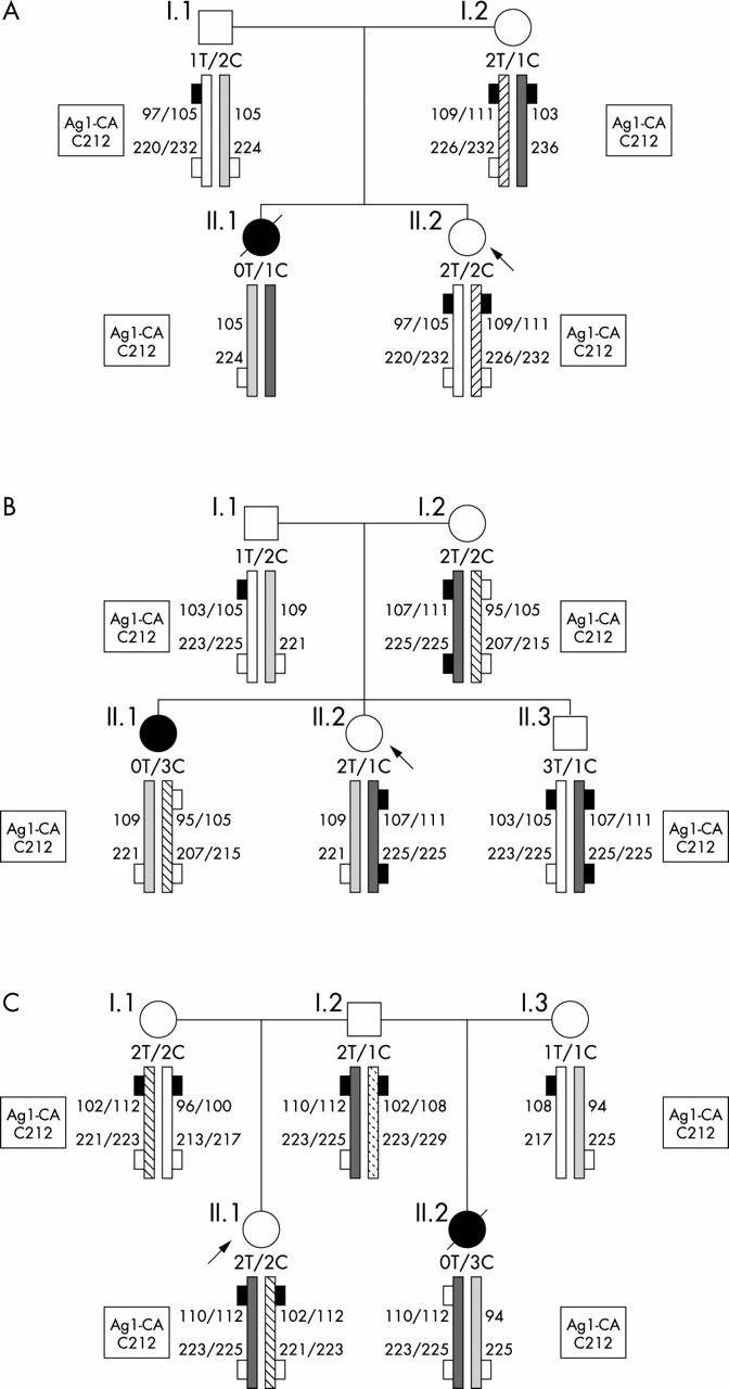
In the parent population, carrier status (only one SMN1 gene detected) was confirmed in 193 (95.5%). Nine families had one parent carrying two SMN1 copies (4.5%). We found two de novo deletions (1.0%), one on a maternally and one on a paternally derived chromosome. Six parents with SMN1 duplication (3.0%) were detected. Finally, one de novo gene conversion on a paternally derived chromosome, converting an SMN1 gene into SMN2, was found (0.5%) in which a normal chromosome carrying one SMN1 and one SMN2 was changed into a pathological chromosome carrying two SMN2 copies. Three examples of the tested families are shown in fig 1A–C.

Three examples of the tested families. The SMN genotype (T for telomeric gene SMN1, C for centromeric gene SMN2) and results of the marker analysis are given above each individual symbol. Patients asking for genetic counselling are indicated with arrows. Full rectangle shows SMN1, open rectangle shows SMN2. (A) Family 1 is an example of de novo deletion. This molecular mechanism could be suspected from the SMN gene dosage alone, mainly because dosage of a single SMN2 in the proband was not coherent with the genotypes of her sister and parents. This result was confirmed by marker analysis, which showed the loss of the maternal contribution. To establish which chromosome was deleted in the affected child, we analysed D5S637 and D5S629, two markers flanking the duplicated region. We found that the deleted chromosome was the one carrying one SMN1 gene. (B) In family 2, an SMN1 duplication was detected in family member I.2. Gene dosage in family member II.2, being tested for carrier detection, showed two SMN1 genes: only by combining results of both analyses (quantitative and marker) in all family members could we show that she was an SMN1 deletion carrier. (C) Family 3 shows gene conversion. In family member II.2, the affected daughter, the five known divergences between SMN1 and SMN2 genes were tested by sequencing and restriction enzyme digestion and all showed SMN2 origin.7 Moreover, she carried three SMN2 genes, whereas her parents each carried only one SMN2 gene. Paternity was confirmed. Analysis of markers C212 and Ag1-CA showed that both daughters had inherited the same chromosome from their father. Taking all these findings together, a de novo conversion of SMN1 into SMN2 was the only mechanism that could account for them. Therefore, we deduced a normal genotype for family member I.1.
Genotypes in the general population
Most of the people from the general population were spouses of relatives with SMA who reported no neuromuscular diseases in their families. Among them, 11 heterozygous genotypes (one SMN1 copy) were detected. These data set the carrier prevalence at 1/34 in the general population (table 1). Moreover, we estimated the prevalence of the genotype with SMN1 duplication and SMN1 deletion by multiplying the two chromosome frequencies as independent probabilities (table 2). About 1/1000 people with two SMN1 genes is expected to carry a duplication plus deletion genotype. This result overlaps with the estimation deduced from our data in the carrier population. Indeed, if 3% of parents show SMN1 duplication and carrier frequency in the general population is 1/34, a duplication plus deletion genotype should be found in 1/1133 people with a normal genotype, including two or more SMN1 genes.
SMN1 genotypes in the general population
Chromosomes in the general population
DISCUSSION
Carrier testing in parents and relatives of patients with SMA raises several problems about genotype interpretation and consequently assessment of recurrence risk. Carrier screening in parents of children with SMA with SMN1 homozygous deletion is the method for detecting atypical genotypes. We found that 4.5% of parents carried two SMN1 genes. We therefore characterised the molecular mechanism to establish the recurrence of risk in these families accurately. In fact, detecting a de novo deletion reduces the risk of recurrence in a family from 25% to less than 1% as germinal mosaicism has never been detected. Conversely, SMN1 duplication is rarely considered when analysing families with SMA, although it is even more frequent than de novo deletion and leads to errors in genotype interpretation in relatives. Finally, conversion of SMN1 into SMN2 confirms that this mechanism induces duplication of SMN genes and de novo loss of the SMN1 gene that is not detected by marker analysis.
When calculating the risk of recurrence in a couple where one is a carrier (one SMN1 detected) and the other has a two SMN1 genotype, three possibilities should be considered: (1) the presence of a subtle mutation in the person with the two SMN1 genotypes (as in 1% of SMA parents7,10 estimated at 1% of 1/34 in the general population); (2) the transmission of a de novo deletion (as in 1% of SMA parents, estimated at 1% of 1/34 in the general population); (3) the presence of a SMN1 duplication associated with a deletion (estimated at 1/1000 of the two SMN1 genotypes in the general population (our study)). Adding the risks derived from the three different molecular mechanisms, we calculate that the risk of recurrence will be 1/2520 for each pregnancy.
Intriguingly, carrier prevalence is higher than previously deduced from the disease prevalence: direct estimation sets carrier prevalence in the general population at 1/34 rather than 1/40. This finding increases the prevalence of SMN1 deleted genotypes to 1/4624. Underestimation of the frequencies of the extreme phenotypes could probably explain this discrepancy. The genotype with complete deletion of SMN1 and SMN2 should exist even if it has never been detected, as it would almost certainly be lethal, maybe at the embryonic stage, leading to early miscarriage. Conversely, non-symptomatic SMN1 homozygously deleted people have already been described.28,29 The question about the predictive value of this finding should be considered along with data concerning the age of evaluation and the clinical course of such patients.29,30 Only a few patients with SMA who have type IV and type III SMA with chronic evolution have been reported8,9 as they often remain undiagnosed because of the delayed onset of the disease and reasonable level of fitness. Further investigations are necessary to determine the exact prevalence of all SMA phenotypes. Studies about the level of expression of SMN2 in these patients will probably elucidate the phenotypic variability in SMA.