Article Text

Abstract
Purpose: To identify the biochemical and molecular genetic defect in a 16-year-old patient presenting with apical hypertrophic cardiomyopathy and neuropathy suspected for a mitochondrial disorder.
Methods: Measurement of the mitochondrial energy-generating system (MEGS) capacity in muscle and enzyme analysis in muscle and fibroblasts were performed. Relevant parts of the mitochondrial DNA were analysed by sequencing. Transmitochondrial cybrids were obtained by fusion of 143B206 TK− rho zero cells with patient-derived enucleated fibroblasts. Immunoblotting techniques were applied to study the complex V assembly.
Results: A homoplasmic nonsense mutation m.8529G→A (p.Trp55X) was found in the mitochondrial ATP8 gene in the patient’s fibroblasts and muscle tissue. Reduced complex V activity was measured in the patient’s fibroblasts and muscle tissue, and was confirmed in cybrid clones containing patient-derived mitochondrial DNA. Immunoblotting after blue native polyacrylamide gel electrophoresis showed a lack of holocomplex V and increased amounts of mitochondrial ATP synthase subcomplexes. An in-gel activity assay of ATP hydrolysis showed activity of free F1-ATPase in the patient’s muscle tissue and in the cybrid clones.
Conclusion: We describe the first pathogenic mutation in the mitochondrial ATP8 gene, resulting in an improper assembly and reduced activity of the complex V holoenzyme.
Statistics from Altmetric.com
Mitochondrial (mt) ATP synthase, or complex V (EC 3.6.3.14), uses the proton gradient provided by the activity of the respiratory chain enzymes complexes I, III and IV for ATP synthesis, thereby generating >95% of cellular ATP.1 Complex V is a multisubunit complex consisting of two functional domains, F1 and F0, connected by a stalk. The F1 domain contains five different subunits (three α, three β, and one γδ∊), is situated in the mitochondrial matrix and acts as the catalytic domain. The F0 domain is embedded in the mitochondrial inner membrane and consists of eight subunits (a–g and A6L). The stalk contains the subunits OSCP, F6, b and d.2 Protons pass from the intermembrane space to the matrix through F0, which transfers the energy created by the proton electrochemical gradient to F1, where ADP is phosphorylated to ATP.23 It has been proposed that a shaft (“rotor” consisting of the ring of 10 c subunits, plus the subunits γ, δ and ∊ that form the central stalk) linked to F0 rotates relative to the catalytic subunits (three α, three β) of F1, thereby sequentially changing the conformation of these subunits, creating the “binding change mechanism of energy coupling” proposed by Boyer.4–6 Two of the F0 subunits, subunit a (or subunit 6) and subunit A6L (or subunit 8) are encoded by the mtDNA ATP6 and ATP8 genes, respectively.7 To date, mtDNA complex V mutations have only been described in the mitochondrial ATP6 gene (MT-ATP6). The point mutations m.8993T→G/C are the most commonly encountered MT-ATP6 mutations and, depending on the degree of heteroplasmy, mainly lead to the clinical picture of NARP (neuropathy, ataxia, and retinitis pigmentosa) or to the more severe MILS (maternally inherited Leigh syndrome).1 8 ATP synthase deficiency due to nuclear genetic defects has also been described.1 9 To our knowledge, pathogenic mutations in the mitochondrial ATP8 gene have not been published to date.10 We describe a 16-year-old patient presenting with apical hypertrophic cardiomyopathy and neuropathy. Biochemical analysis performed in muscle tissue and fibroblasts showed an isolated complex V deficiency. Mutation analysis of mtDNA revealed a pathogenic nonsense mutation in MT-ATP8. We studied the molecular pathophysiology of this novel mtDNA mutation using transmitochondrial cybrid cells.
CASE REPORT
The patient is the third child of healthy, non-consanguineous Caucasian parents. His older brother and sister are healthy. The family history is unremarkable. According to the parents, his speech development was delayed but turned to normal after an adenotomy at the age of 4 years. He has worn glasses since the age of 3 years for hypermetropia and astigmatism, and one eye was temporarily covered because of amblyopia. His motor development has been slower than his siblings since infancy. He walked independently at the age of 18–24 months, and learned to ride a bicycle at the age of 7–8 years. During childhood, he had frequent falls, and his gross motor skills were clumsy. At the age of 9–10 years, his balance problems became prominent. Since then there have been reports of reduced muscle strength, walking problems and tingling in the limbs. These symptoms appear to be progressive. Exercise intolerance, shortness of breath (dyspnoea) during exercise, and angina were also common symptoms.
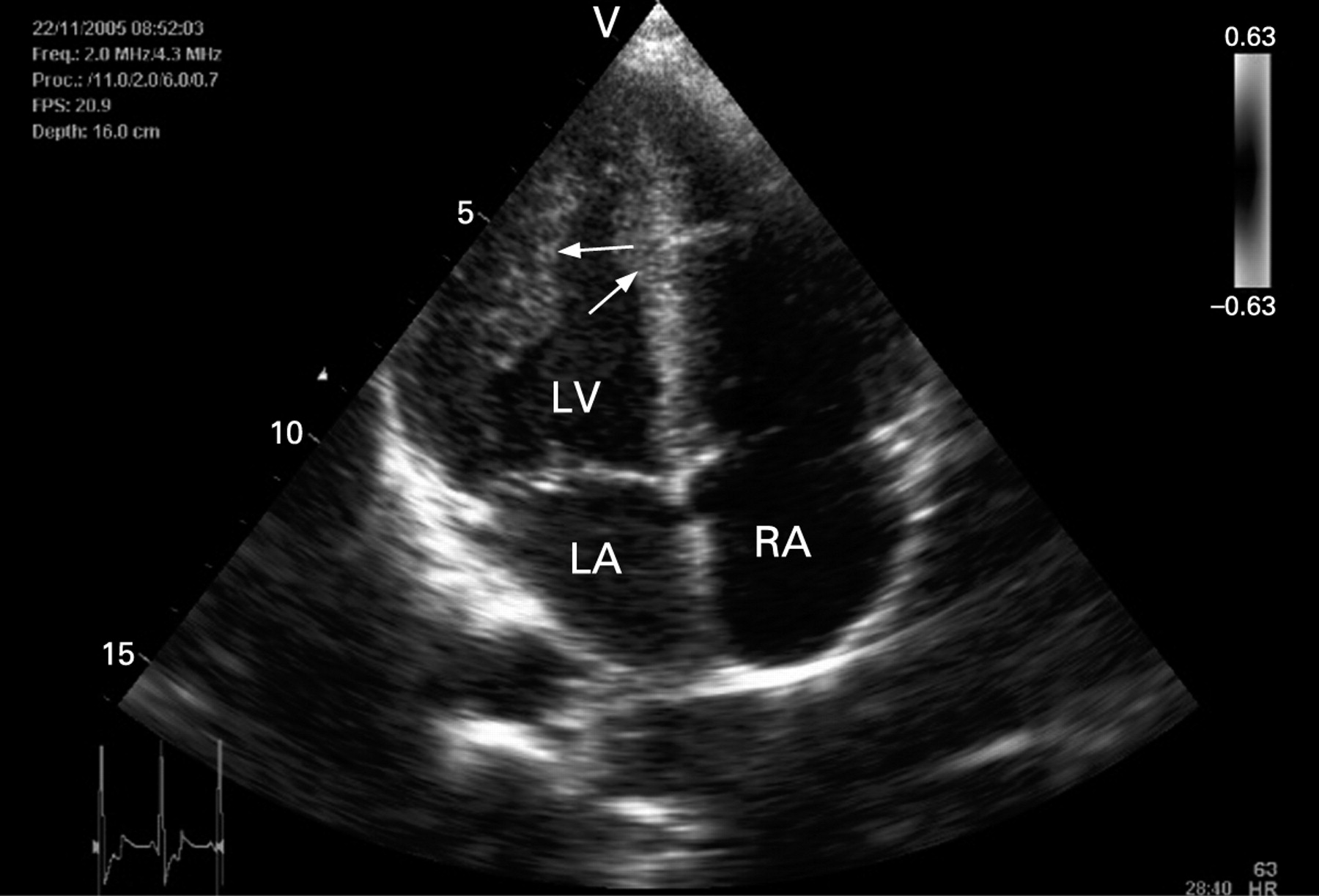
Clinical examination at the age of 16 years showed a slender young man, whose speech was dysarthric. He had an ataxic walking pattern and revealed a clear Trendelenburg sign. Reduced tendon reflexes and a bilateral Babinski sign were present. Moderate external ophthalmoplegia was noted. Metabolic tests performed at the age of 14 years showed normal blood lactic acid concentrations, with increased lactic acid in the cerebrospinal fluid (4000 µmol/l; normal range 1350 to 1900). The organic acid pattern in the urine was normal. Ophthalmological evaluation confirmed the astigmatism and hypermetropia, and showed no signs of retinitis pigmentosa. Audiology was normal. MRI of the brain did not show any abnormalities other than a cisterna magna. Neuropsychological evaluation revealed a moderate learning disorder. Cardiac evaluation showed an extensive left ventricular hypertrophy on the ECG, without signs of arrhythmia. Echocardiography was abnormal: left ventricular hypertrophy was concentrated around the apex of the left ventricle (fig 1) with discrete asymmetrical hypertrophy of the echogenic interventricular septum and without left ventricular outflow tract obstruction. A small atrial septal defect with left to right shunt was detected. At this age, sensory and motor axonal polyneuropathy was seen on an electromyogram; the sensory potentials could not be evoked in the sural nerve bilaterally, and there were slightly delayed motor nerve conduction velocities with reduced amplitudes in the peroneal and tibial nerves. Finally muscle (quadriceps femoris) and skin biopsies were taken.

METHODS
Cell cultures
Fibroblasts were cultured in M199 medium (Gibco, Invitrogen, Breda, The Netherlands) supplemented with 10% fetal calf serum and penicillin/streptomycin (respectively 100 U/ml and 100 µg/ml). Transmitochondrial cybrids were obtained by fusion of 143B206 TK− rho zero cells with enucleated fibroblasts derived from patient or control as described previously.11 Seven independent colonies (clones) of the patient transmitochondrial cybrid cell line were randomly picked for further study. For the control cybrid cell line, the colonies were pooled, resulting in a clone mixture.
Biochemical assays
Measurement of MEGS capacity was performed as described previously.12 The complex V (or mtATPase) activity was measured spectrophotometrically in mitochondria isolated from frozen pellets of the patient and control cybrids as described previously.8 Briefly a 980 μl solution containing 250 mmol/l sucrose, 50 mmol/l KCl, 50 mmol/l K2HPO4, 50 mmol/l KH2PO4, 1 mmol/l phosphoenolpyruvic acid, 11 nmol/l P1-P5-di(adenosine-5’) pentaphosphate, 0.2 mmol/l EGTA, 5 mmol/l ATP, 5 mmol/l MgCl2, 0.2 mmol/l oubaine, 2 μmol/l carbonyl cyanide 3-chlorophenyl hydrazone (CCCP), 250 μmol/l NADH, 1.25 μmol/l rotenone, 2.5 U/ml lactate dehydrogenase, and 1.5 U/ml pyruvate kinase was incubated for 10 minutes at 30°C. A mitochondrial suspension was freeze-thawed three times and 20 μl was added to the reaction mixture, mixed and transferred to a cuvette, after which the absorption at 340 nm was measured for 8 minutes. Subsequently, 1 μl of an 8 mg/ml oligomycin solution in ethanol was added and the absorption was measured for another 8 minutes. The oligomycin-sensitive activity of complex V was calculated using an ∊340 for NADH of 6.22×10³ l/mol×cm and was expressed in units (the amount of enzyme required to convert 1 µmol NADH/min) per unit cytochrome oxidase (COX)13 and the mitochondrial matrix enzyme citrate synthase (CS)14 activities, respectively. The activities of the mitochondrial respiratory chain enzymes were measured spectrophotometrically in muscle tissue and fibroblasts of our patient according to described protocols.15 16
Molecular genetic analysis
Total DNA from frozen skeletal muscle tissue and from cultured fibroblasts was extracted using the Gentra genomic isolation kit according the protocol of the manufacturer (Biozym, Landgraaf, The Netherlands). Major DNA deletions or other rearrangements in the mtDNA were screened for using standard long-template PCR. The presence of the MELAS m.3243A→G, MERFF m.8344A→G and Leigh/NARP m.8993T→G/C point mutations was investigated using Pyrosequencing technology as described previously.8 17 Sequence analysis of PCR amplified products of the mtDNA-encoded tRNAGln, tRNAGly, tRNAIle, tRNALeu(cun), tRNALeu(uur), tRNALys, tRNASer(agy) and mitochondrial ATP6 and ATP8 genes was performed on an ABI3730 automatic capillary sequencer using BigDye terminator chemistry (Applied Biosystems, Nieuwerkerk a/d ljssel, The Netherlands). Primers used for amplification of tRNA genes are available upon request. MT-ATP6 and MT-ATP8 were amplified by PCR as three overlapping fragments (a, b and c) using the primers shown in table 1. PCR conditions were 92°C for 30 seconds, 55 °C for 30 seconds and 72°C for 60 seconds, for 35 cycles. Subsequently, mtDNA was analysed in the patient and control cybrids looking for the nonsense mutation m.8529G→A following the protocol described above.
Complex V assembly and activity
Blue native polyacrylamide gel electrophoresis (BN-PAGE), blotting and measurement of complex V in-gel activity was performed as described previously,18 19 loading 60 μg of protein20 of oxidative phosphorylation complexes isolated from a mitoplast fraction of the patient and control cybrids and muscle homogenates. For Western blotting, monoclonal antibodies against mitochondrial ATP synthase subunit α and CoII-70 kDa (complex II) (Invitrogen, Breda, The Netherlands) were used. For the complex V in-gel activity assay the gel was incubated overnight at room temperature with the following solution: 35 mmol/l Tris, 270 mmol/l glycine, 14 mmol/l MgSO4, 0.2% Pb(NO3)2, and 8 mmol/l ATP, pH 7.8.19
RESULTS
Biochemical assays
Measurement of MEGS capacity in muscle tissue showed a reduced pyruvate oxidation rate, which improved after addition of CCCP, pointing to a possible complex V defect.12 The ATP production rate from oxidation of pyruvate was reduced. Spectrophotometric analysis confirmed the reduced complex V (mtATPase) activity in muscle tissue and in fibroblasts. The activities of the respiratory chain enzymes and CS were normal (table 2).
Molecular genetic analysis
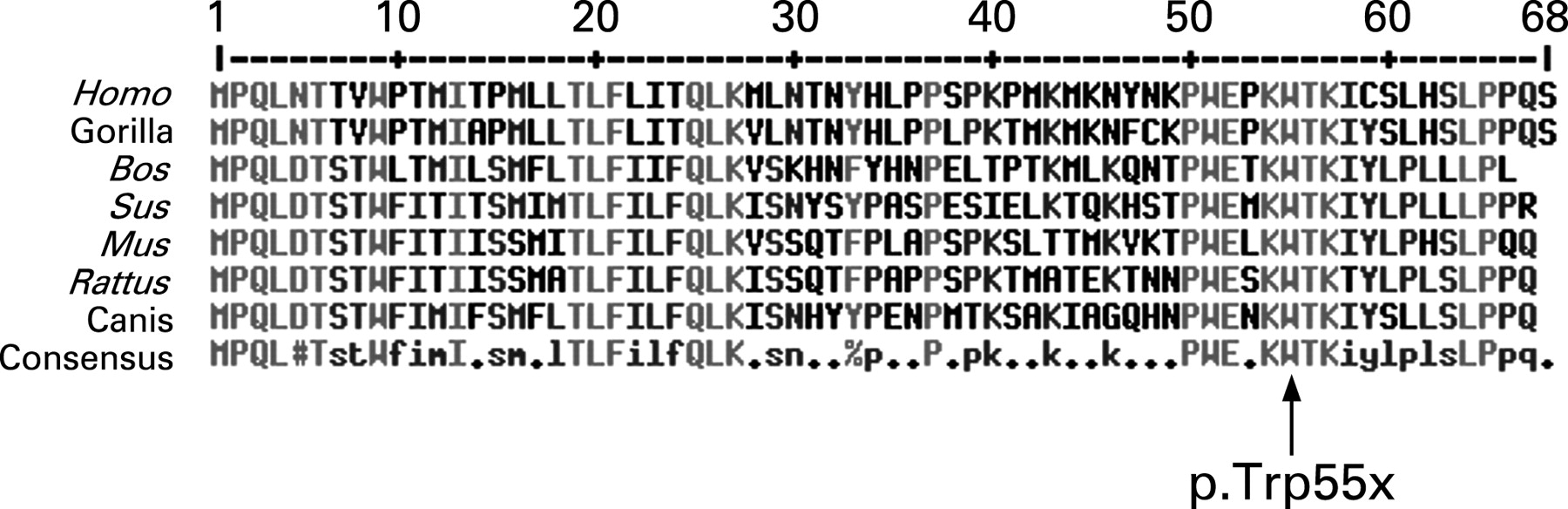
The diagnosis of a mitochondriopathy was further substantiated by mutation analysis of mtDNA from muscle tissue and fibroblasts, showing a homoplasmic (→90%) nonsense mutation m.8529G→A (p.Trp55X) in the mitochondrial ATP8 gene, located in an overlap region of MT-ATP6 and MT-ATP8 (nucleotides m.8527–8572). It results in a silent change in MT-ATP6 (Met1Met; ATG→ATA), whereas it introduces a premature stop codon in the C-terminal domain of MT-ATP8, which is a conserved region 10 21 (fig 2). This probably results in a truncated protein lacking the last 14 amino acids.
Cell biological consequences
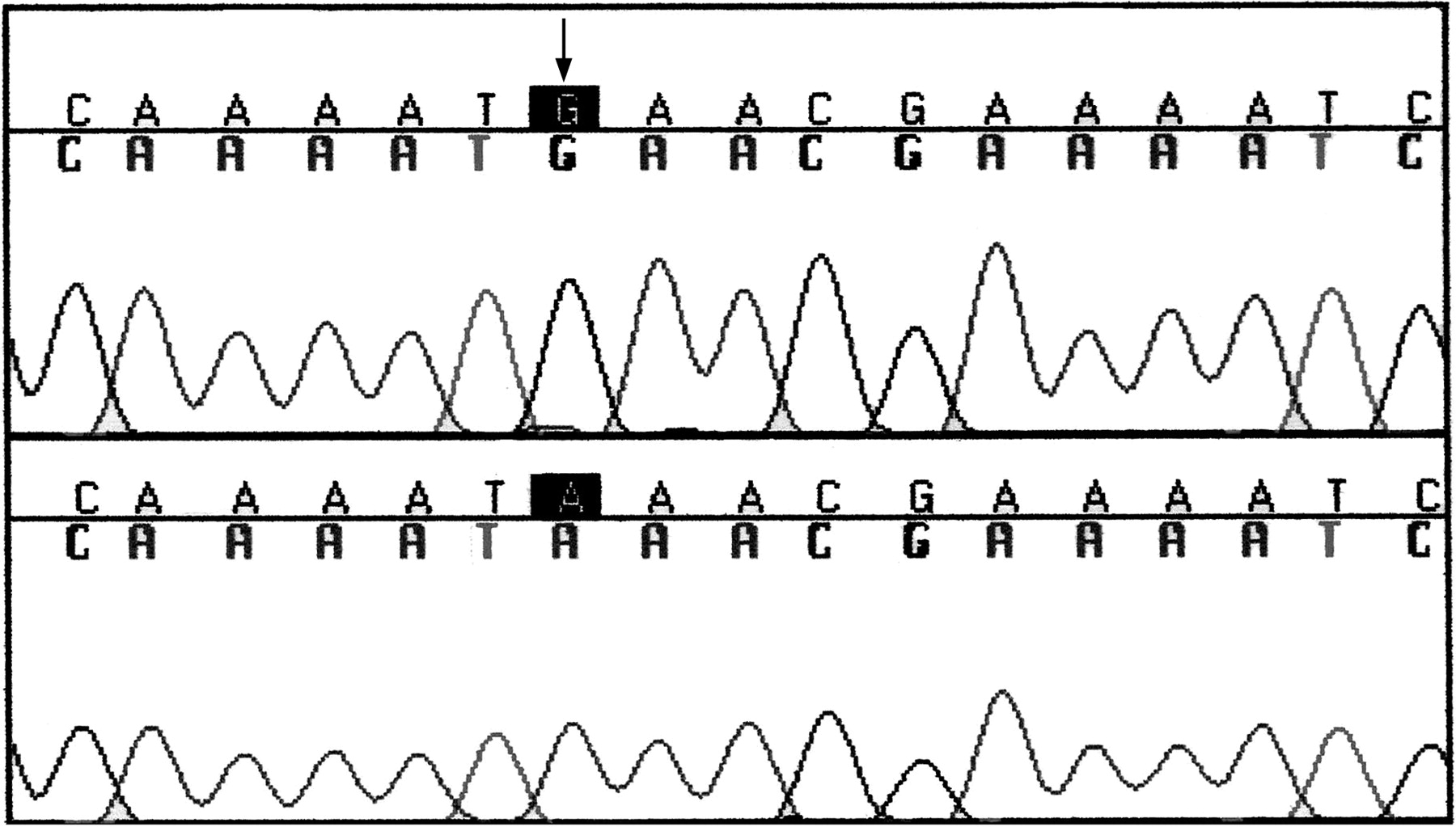
DNA analysis in the cybrid cells confirmed the presence of the mitochondrial nonsense mutation m.8529G→A in each of the clones containing patient-derived mtDNA. Sequence analysis showed that all clones were homoplasmic for the mutation. The control cybrid cell line showed no mutation (fig 3).
Enzymatic analysis of complex V was performed in the patient cybrid clones and in the control cybrid clone mixture. A distinctly reduced activity was found in the patient cell line relative to the control (table 3). CS and COX activities were normal in the patient and control cybrids (data not shown).
A larger range of residual activities in individual cybrids is seen when normalised to the COX activity because the COX assay has a broader normal range than the CS assay.
Immunoblotting after blue native polyacrylamide gel electrophoresis (fig 4) with anti-ATP synthase subunit α antibody clearly revealed a lack of holocomplex V and increased amounts of subcomplexes of mitochondrial ATP synthase in the patient cybrid clones and muscle homogenate. This was not found in the control cybrid and control muscle, which showed only the complex V holoenzyme and dimer. The most likely subcomplexes are the F1-ATPase and the subcomplex V*, as denoted previously.2 22 The in-gel activity assay of ATP hydrolysis showed activity of free F1-ATPase in the patient cybrid clones and muscle. The holoenzyme showed no activity in either the patient or control samples (fig 5).


DISCUSSION
Clinical phenotype
We describe the first pathogenic mutation in the mitochondrial ATP8 gene ,to our knowledge. It has been stated previously that the clinical phenotype of complex V deficiency due to nuclear genetic defects (hypertrophic cardiomyopathy, hypotonia, facial dysmorphism and microcephaly) markedly differs from the severe central system changes observed in NARP or MILS.1 The clinical phenotype of our patient is characterised by signs and symptoms that have mainly been described both in mitochondrial (neuropathy, ataxia) and nuclear (hypertrophic cardiomyopathy) complex V defects. Hypertrophic cardiomyopathy can present with negligible to extreme hypertrophy, minimal to extensive fibrosis and myocyte disarray, absent to severe left ventricular outflow tract obstruction, and distinct septal contours/morphologies with extremely varying clinical course.23 The apical variant hypertrophic cardiomyopathy reported here represents a genotype–phenotype relationship that has not been described previously. Taken together, these data indicate that the clinical variability among the patient group carrying mitochondrial complex V mutations is greater than previously assumed.
Pathogenicity
Our results strongly suggest that the MT-ATP8 mutation is the cause of the complex V deficiency in our patient. Several lines of evidence point to this: (1) the mutation leads to a premature stopcodon situated in the C-teminal region of subunit 8; (2) the C-terminal domain of mitochondrial ATP synthase subunit 8 is a conserved region in yeast21 and mammals10 (fig 2); (3) no polymorphisms have been described previously at the position of this mutation;10 24 (4) the mutation is homoplasmic (>90%) in muscle tissue and fibroblasts, and in both tissues the complex V activity is clearly reduced (table 1); and (5) transmitochondrial cybrids retained the complex V deficiency, confirming the mitochondrial origin of the pathogenic mutation.
Molecular pathophysiology
Immunoblotting after BN-PAGE showed a lack of holocomplex V and increased amounts of subcomplexes, and the in-gel activity assay found isolated activity of F1-ATPase in the cybrids and in muscle. The results of the in-gel activity assay of ATP hydrolysis probably point to the free rotation capacity of the F1 subcomplex in the patient. It was reported that F1 can be detected as a single entity2 and that it retains ATP hydrolysis activity.22 The higher in-gel ATP hydrolysis activity of F1-ATPase has been described previously.22 It has been suggested that subunits of the F0 portion of ATP synthase restrict in-gel ATP hydrolysis,22 which results in an apparently low activity of the holocomplex in this assay. Whereas the in-gel activity assay showed an isolated F1-ATPase activity, the spectrophotometric assay demonstrated a very low F0F1-ATPase activity.
The enzymatic assay measures the oligomycin-sensitive activity of complex V.8 This is the result of the subtraction of the mtATPase holoenzyme activity and the activity after addition of oligomycin, an inhibitor of proton translocation in mtATPase.25 Athough the ATPase activity in the absence of oligomycin was of the same order of magnitude in our patient as in controls, the activity in the patient was not sensitive to oligomycin inhibition, whereas in control cells, usually a 90% inhibition is seen. In fact, detachment of the F1 from the F0 subcomplex, as seen on the BN gel data of the patient cybrid clones and muscle homogenate, leads to a decrease in oligomycin-sensitive complex V activity. ATP hydrolysis by F1 takes place, but without the coupling of ATP hydrolysis to extrusion of protons through F0, the sensitivity to oligomycin is no longer evident.25
How can the increased amounts of subcomplexes of complex V be explained? It has been reported that subunit 8, which is unique to the mtATPase of fungi and mammals, consists of three functional domains: the N-terminus, a central transmembrane hydrophobic domain and the C-terminus localised to the mitochondrial matrix side.26 27 In yeast, the positively charged C-terminal region has been shown to be required for assembly of yeast subunit 8 (Y8) into the F0 sector.28 As Y8 interacts with other subunits in the assembly of the F0 sector, subunit 6 assembly into mtATPase requires the presence of assembled Y8 in the enzyme complex,29 thus it could be speculated that mutations in the C-terminal region, as present in our patient (the p.Trp55X, subunit 8 having 68 residues) are associated with reduced complex V assembly, which appears to be similar to the effect of human MT-ATP6 mutations.22 Our immunoblotting results confirm this hypothesis.
The impaired complex V assembly very likely has a repercussion on the function of the holoenzyme, as was shown in our patient by enzymatic analysis. Impaired subunit 6 assembly into the F0 sector possibly plays a role. It has been reported that subunit 6 has two functions: first, it leads the protons through a hydrophilic half-channel into the matrix, and second, it turns the rotor for ATP synthesis.30 The MT-ATP6 m.8993T→G mutation interferes with the rotation of the ring of c subunits, and therefore it uncouples ADP phosphorylation in F1 from proton transfer through F0.30 The assembly problem is probably not the sole pathogenic factor. It is also likely that the subunit 8 protein is reduced due to the nature of the mutation in our patient, namely a premature stop codon, probably resulting in a truncated protein. In that case, a direct negative effect on the function of the enzyme could be assumed. It has indeed been described in yeast that Y8 also plays an important role in determining the mtATPase activity.26 In particular, the hydrophobic nature of amino acids in the centre of the transmembrane domain of Y8 is essential for coupling proton transport through F0 to ATP synthesis on F1.26 Thus, Y8 may take part in conformational changes that occur between the F0 and F1 sectors of the enzyme during catalysis.26 However, further investigation is needed to confirm this.
Acknowledgments
AIJ and JS are supported by a grant from the Prinses Beatrix Fonds.
REFERENCES
Footnotes
Competing interests: None.